Skip to content Skip to sidebar Skip to footer

HAND FRACTURE

Definition

A hand fracture is a partial or complete break in one or more of the bones that make up the structure of the hand. The hand is made up of several bones, including the metacarpals (the bones between the wrist and fingers) and the phalanges (the bones of the fingers). Hand fractures can occur as a result of direct trauma (such as a blow or fall), indirect trauma (such as a sudden movement), or impact to the hand.

Symptoms of a hand fracture can include pain, swelling, deformity, tenderness, bruising and difficulty moving the hand or fingers. The diagnosis of a hand fracture is usually confirmed by X-rays, which help assess the location and severity of the fracture.

Treatment of a hand fracture will depend on a number of factors, including the type of fracture, its location, and the presence of any bone displacement. In some cases, simple immobilization with a splint or cast may suffice to allow the fracture to heal. In other cases, surgery may be required to realign and fix the fractured bones with screws, plates or other devices.

Recovery from a hand fracture can vary depending on the severity of the fracture and the treatment applied. Rehabilitation may be required to restore hand mobility and strength after healing.

What are the symptoms?

On consultation, the characteristic symptoms are often as follows: the patient describes a feeling of discomfort or pain of varying severity; physical examination also reveals inflammation, altered shape, haematoma, limited functionality (difficulty or inability to grasp or open the hand) and pain when pressure is applied to the bone.

What tests should be carried out in the event of a fracture?

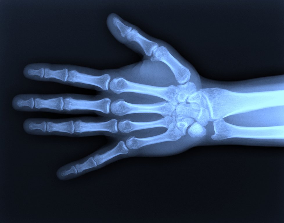
X-rays of the affected hand are the main examination performed. It enables the precise severity of the injury and the number of bones involved to be determined. If the practitioner suspects damage to the scaphoid bone, or if the hand shows previous fractures with deformity or non-union, additional examinations such as CT scan, scintigraphy or MRI may be carried out.
How is a hand fracture treated?

The aim of the treatments administered is to realign the fracture in order to promote rapid and complete recovery of hand mobility.

Orthopedic treatment

This treatment involves appropriate immobilization for slight or non-displaced fractures. You will be monitored regularly with X-rays to ensure that no displacement of the fracture occurs. Immobilization usually lasts between 3 and 6 weeks, after which it may be replaced by syndactyly (grouping 2 or 3 fingers together with a bandage) to facilitate finger mobilization.

surgical treatment

For displaced, multiple or unstable fractures, a surgical procedure called osteosynthesis is required. This procedure is performed under locoregional anesthesia. First, the fracture is realigned using X-ray guidance. Next, the surgeon will fix the fracture using osteosynthesis hardware, such as pins, plates or screws, to promote the natural healing of the bone.

Wires and external fixators are usually removed in the operating room around 6 weeks after surgery. Screw plates remain in place for around 1 year, before being removed if necessary. In older patients, the hardware may be left in place for life, unless it causes discomfort, in which case the patient may request its removal.

A plaster cast or resin can be used temporarily to provide additional support.

As with any surgical procedure, this treatment entails surgical risks, some of which are general and others specific:

  • A local or generalized infection, the severity of which depends on when it is treated.
  • Failure of bone consolidation, with smoking as an unfavourable factor.
  • Maintaining stiffness in the hand.
  • Subsequent fracture displacement.

In addition, there is a risk of algodystrophy, a condition characterized by swelling, pain, excessive sweating and stiffness of the hand. Although rare, this can be a worrying development, as it can lead to residual pain and stiffness. It may persist for several months.

What happens after the operation?

After surgery, careful clinical follow-up is planned to monitor healing and detect any signs of post-operative infection. Appointments are scheduled at specific intervals, usually 2 to 3 weeks after surgery, then at 6 weeks to 2 months, and finally at 3 to 6 months. At these appointments, X-rays will be taken to check that there has been no displacement of the fracture and to assess bone consolidation. These X-rays can be taken in the same department on the day of the surgical consultation.

A nurse will change the dressing every other day until the fracture is completely healed. Your hand will be immobilized for between 1 and 3 months, depending on the type of fracture. Physiotherapy sessions will be prescribed to facilitate rehabilitation. You will be able to resume sporting activities after 1 to 2 months following the end of immobilization, and this may require the use of an orthosis.

Upload Files Drag and drop your files here or click the button to upload your files from your computer

multi file upload Drag and drop your files Max Upload: 20 MB
close image preview
image preview
Task already exist for this element.
Write your message in the existing thread.
Here, we opened it for you.
Pushed to Media Folder.
The file was added to the website's media folder, you can now use it from the there.
poweredby
hide sidebar icon
sidebar left
sidebar menu
sidebar menuThis Page
sidebar menuAll Pages
sidebar menuShow Complete Tasks
sidebar menuShow Internal Tasks
sidebar menuSort by Date
sidebar menuSort by Priority
sidebar menuSort by Status
Tasks
Pages
no task in sidebar
Add your comments
Click any part of the page to start collaborating
              Loading...
              ×
              Approve Page

              When the page is ready, click the green button to signal to the webmaster that <b>this page is approved.

              there was some error. Please try again.
              ×
              Page Approved
              Desktop Tablet Mobile
              Back to Standard View
              You Ran Out of Website Slots

              That's great!
              It means you're growing & onboarding more clients!<br>Don't let it stop you

              There was some error. Please try again.

              Tendonitis Hand Wrist

              Tendonitis is an acute or chronic inflammation of the tendons, often involving their anchoring points on the bones (apophysitis), their synovial sheaths (tenosynovitis) or their bursae (tenosynovitis)….

              Osteoarthritis of the wrist

              Osteoarthritis of the wrist is a condition in which the wrist joints are affected by progressive deterioration of the cartilage covering the joint surfaces. This deterioration can lead to pain, stiffness, reduced mobility, …

              Joint cysts

              It’s an abnormal mass that can develop in different places on your hand. The most common position is on the back of the wrist. However, it can also appear at the front of the wrist, or more rarely at the base of a finger.

              Osteoarthritis of the fingers

              Osteoarthritis of the fingers, also known as digital osteoarthritis, is a degenerative condition affecting the finger joints. It is characterized by the progressive degradation of the cartilage that covers the ends of the joint bones.

              Finger locks

              Finger locking, also known as finger springs or spring fingers, is a medical condition where a finger or thumb becomes difficult to bend or unfold due to a sudden, uncontrollable contraction of the flexor tendons.

              Carpal tunnel - Median nerve compression

              What is the median nerve? The median nerve is one of the major nerves of the hand and arm. It originates in the spinal column and runs the length of the arm to reach the hand.

              Ulnar wrist pain

              Ulnar wrist pain, also known as ulnar pain, refers to a sensation of discomfort or pain felt on the ulnar or medial side of the wrist, i.e. on the side of the little finger.

              Dupuytren's disease

              Dupuytren’s disease is characterized by the formation of fibrous tissue (also known as fibromatosis) under the skin of the hand, leading to gradual contraction of one or more fingers towards the palm. The origins of this condition are many and varied, including familial predisposition.

              Osteoarthritis of the thumb

              Osteoarthritis of the thumb, also known as rhizarthrosis, is a degenerative joint condition that involves the progressive degradation of cartilage at the base of the thumb, where the wrist bone called the trapezium meets the first bone of the hand, the metacarpal. This cartilage degradation can lead to pain, stiffness and reduced joint function in this area.

              Dupuytren's disease

              Dupuytren’s disease is characterized by the formation of fibrous tissue (also known as fibromatosis) under the skin of the hand, causing gradual contraction of one or more fingers towards the palm. The origins of this condition are many and varied, including familial predisposition. However, despite its first description by Baron Guillaume Dupuytren in 1832, the precise origin of the disease remains unknown.

              Our surgeon makes you feel more confident.

              Lorem ipsum dolor sit amet, consectetur adipiscing elit. Aenean dictum dolor nunc, at consectetur nulla volutpat ut. Nam suscipit ex sapien, et vehicula lectus dapibus imperdiet. Curabitur ac metus magna. Nunc sagittis justo a nunc vulputate, in euismod quam aliquet. Donec non hendrerit sapien.